imag-alam-04-09-18-1200x334.jpg

Septiembre 4, 2018

La inmensa y prolífica carrera del Dr. Romilio Espejo ha sido justamente reconocida esta semana con el Premio Nacional 2018 de Ciencias Aplicadas y Tecnológicas. El Dr. Espejo, quien fue uno de los primeros egresados de la carrera de Bioquímica de la Facultad de Ciencias Químicas y Farmacéuticas de la Universidad de Chile, se desempeña actualmente como investigador del Instituto de Nutrición y Tecnología de los Alimentos (INTA) de esta misma casa de estudios y es Socio de la Sociedad de Microbiología de Chile (SOMICH). El Premio reconoce su contribución en el desarrollo de la biología molecular y epidemiología tanto a nivel bacteriano como viral, como también sus aportes en el área de la biominería. El académico de la Universidad de Chile había sido también reconocido previamente por la Academia Americana de Microbiología (1999) y nombrado Miembro de Número de la Academia Chilena de Ciencias del Instituto de Chile (2013).

En nombre del Directorio de la SOMICH y todos/as nuestros/as socios/as queremos entregar las mas sinceras felicitaciones a nuestro colega y amigo Dr. Romilio Espejo.
http://www.uchile.cl/noticias/146482/romilio-espejo-gano-el-premio-nacional-de-ciencias-aplicadas


img-destacada-alam-1200x334.jpg

Agosto 20, 2018

La Sociedad Española de Microbiología (SEM) en conjunto con la Sociedad de Microbiología de Chile otorgaron 30 becas de inscripción para el Congreso ALAM 2018. Para hacer efectiva esta beca deberá acercarse al stand de inscripción durante el Congreso, donde se reembolsará el precio de la inscripción equivalente del que usted pagó en pesos chilenos.

Esperamos que disfrute su participación en este congreso, donde se reunirán más de 1200 asistentes de América Latina,  Europa y Norte América, que podrán compartir sus experiencias y generar lazos de colaboración.

Revisar ganadores/as.

 Brenda Gómez Ortiz  México
 Carolina F. Cubillos Leòn  Chile
 Chantal L. Márquez  Australia
 Daniela Arturo Terranova  Colombia
 David Rodrigo Cajas Muñoz  Chile
 Edwin Barrios Villa  México
 Erasmo Gámez-Espinosa  Argentina
 Francielly Cristina Machado  Brasil
 Francisca Marcela Oliva Godoy  Chile
 Gabriela Pacheco Sánchez  Brasil
 Geyse Aparecida Cardoso dos Santos  Brasil
 Jeny Patricia de Haro Acosta  México
 Jéssica Ferreira Mafra  Brasil
 Jose Antonio Blanco Mendoza  Chile
 Lidia Nuñez  Argentina
 Lucía Araújo Pírez  Uruguay
 Macarena Victoria Echeverría Bugueño  Chile
 Magalí Andrea Fernández Trinidad  Uruguay
 Manuel Del Cogliano  Argentina
 Maria Elena Bello-Lopez  México
 María Inés Isidro Coxca  México
 Maria Letícia Duarte Lima  Brasil
 Miguel Andrés Mansilla  Chile
 Natalie Castillo Prado  Chile
 Nayara Alves Reis  Brasil
 Tamara Iglesias Sánchez  Uruguay
 Tamara Renata Machado Ribeiro  Brasil
 Thaís Mitsunari  Brasil
 Valentina Carrasco Muñoz  Uruguay
 Victor Andres Sanchez Rivera  Chile

img-alam-1200x334.jpg

Agosto 6, 2018

Tenemos el agrado de informarles que el Comité Organizador del XXIV Congreso Latinoamericano de Microbiología en conjunto con la Sociedad Española de Microbiología-SEM, otorgará 30 becas para participar en este evento.


img-destacada-alam-1200x334.jpg

Julio 18, 2018

El pasado jueves 28 de junio, mas de 60 socios de la SOMICH se reunieron en una cena de camaradería en el Hotel Ritz Carlton, lugar escogido para realizar la Cena-Fiesta de Cierre de ALAM 2018 el próximo 16 de noviembre. En esta oportunidad, los asistentes recibieron información fundamental de la gran responsabilidad que tiene nuestra Sociedad como anfitrión y organizador del Congreso ALAM 2018, así como también aprovechar la oportunidad para recordar que este año habrá elecciones de Directorio 2019-2020. Esta actividad se hará extensiva a otras ciudades del país, como por ejemplo en Antofagasta el próximo 31 de agosto (lugar por confirmar). La idea de estas reuniones es recoger la opinión de los socios, promover la participación y la camaradería.


img-destacada-1200x334.jpg

Mayo 29, 2018

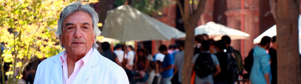
Inducción de la enzima hemoxigenasa-1, que previene el daño en las vías respiratorias ocasionadas por la inflamación, podría ser utilizada como terapia. Director del Instituto Milenio de Inmunología e Inmunoterapia, IMII, dirige estudios sobre este virus, que es principal causa de infecciones severas del tracto respiratorio inferior en niños.

El desarrollo de nuevas drogas antivirales profilácticas y terapéuticas contra el virus respiratorio sincicial (VRS) es fundamental para controlar la carga de enfermedad en la población susceptible. Por ello, el Dr. Alexis Kalergis, académico de la Universidad Católica de Chile y director del Instituto Milenio de Inmunología e Inmunoterapia, IMII, examinó los efectos de una enzima, llamada hemoxigenasa-1 (HO-1),  en la inflamación pulmonar inducida por este virus.“Los resultados de nuestros estudios muestran que después de la infección por VRS, la aplicación de HO-1 disminuyó la replicación viral e inflamación pulmonar de los modelos de análisis transgénicos. Además, observamos efectos antivirales y protectores similares. Finalmente, los datos in vitro sugieren que la inducción de esta enzima puede modular la susceptibilidad de las células a esta infección, especialmente las de tipo epiteliales, de las vías respiratorias”, señaló el académico.

Esta investigación puede complementar a la vacuna contra la enfermedad, desarrollada en nuestro país por el bioquímico, que se ha posicionado a nivel mundial con sus investigaciones  y resultados favorables frente a esta afección, una de las que provoca mayor hospitalización y fallecimiento en menores de dos años. La aplicación de su antídoto ha mostrado seguridad y entregado resultados exitosos en un primer grupo de voluntarios.

La enzima se encuentra de forma natural en el organismo, expresada en muchas células y tejidos, sobretodo en el bazo, riñón e hígado. Sin embargo, también han descubierto que en ciertas patologías, sus niveles están disminuidos. Por esta razón, han podido explicar que la deficiencia de HO-1 en el organismo se asocia a un perfil “inflamatorio importante, y el desarrollo de patologías inflamatorias y otras de carácter autoinmune, como diabetes tipo I y Lupus”, afirma el Dr. Kalergis.

Los científicos del IMII llevan más de una década explorando la acción terapéutica de HO-1. Al respecto, el Dr. Kalergis señala que la enzima tiene capacidad para controlar la función de las células dendríticas, las cuales se encuentran desreguladas en pacientes con problemas de autoinmunidad, promoviendo una sobrerreacción en la respuesta inmune.  ¿Cuál es el secreto de la enzima?  Lo novedoso es que ésta libera pequeñas dosis de un gas responsable de la función antiinflamatoria. “Este gas funciona como un inductor de tolerancia. Y no es tóxico, ya que se libera en cantidades reducidas al interior de las células, y no a nivel sistémico.  Su acción se da en la mitocondria, estructuras que controlan la cantidad de energía, y al hacerlo,  las células bajan su nivel energético, lo que a su vez disminuye la actividad inflamatoria, fomentando así la prevención”, comenta el científico.
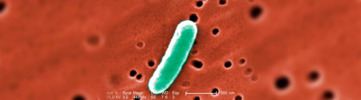
Virus respiratorio

El virus respiratorio sincicial, es de alta incidencia en todo el mundo. Principalmente en los inviernos y favorecido por el frío, contaminación y humedad, este microorganismo ocasiona bronquitis obstructiva, infecciones de vías respiratorias altas, así como neumonía en los casos más severos.

En Chile, el Estado invierte sobre 10 mil millones de pesos anuales en tratamientos, hecho que se suma al colapso de los sistemas hospitalarios. Por estas razones, el doctor en microbiología e inmunología estima que contar con la vacuna en el mercado “será de gran impacto en la comunidad afectada, en sus familias y también traerá beneficios desde el punto de vista económico, ya que permitirá prevenir los daños”.

Considerando que la mayor vulnerabilidad de contagio por el virus, ocurre entre los 0 y 2 años, la estrategia es poder aplicar el antídoto a las pocas horas de nacimiento del bebé. “El blanco principal serán los infantes y pensamos que con una sola dosis bastaría. Nuestra intención es llegar a reemplazar la actual vacuna de BCG, contra la tuberculosis y que la nuestra genere protección contra ambos patógenos”, señala el bioquímico de la Universidad Católica.

Patentes internacionales

En 2017, la vacuna recibió la concesión de la patente china. El profesor titular de la Universidad Católica de Chile, celebró este vínculo con el país asiático, gracias al cual “la vacuna ya cuenta con protección intelectual en esta nación, lo que abre paso a su comercialización y uso en beneficio de millones de personas”.

El antídoto también fue patentado en Estados Unidos el año 2013, con apoyo de un proyecto FONDEF-Interés Público, adjudicado por el científico y su laboratorio “Este hecho representa una contribución importante a resolver un problema de gran significancia para la salud pública mundial”, comenta el Dr. Kalergis.

Su trabajo está orientado a nivel molecular en reestablecer el equilibrio inmunológico en fenómenos autoinmunes. Gracias a la generación de la vacuna contra el virus respiratorio sincicial ha recibido reconocimientos internacionales como la Medalla de Oro para inventores que entrega la Organización Mundial de la Propiedad Intelectual (OMPI). El galardón, el cual se entrega desde 1979, destaca la importancia de los más de diez años de estudios del profesor titular de la Universidad Católica en relación a encontrar una vacuna para el VRS.

Además, realiza investigaciones enfocadas al entendimiento de los mecanismos moleculares responsables de la regulación de la sinapsis inmunológica y desarrollo de enfermedades autoinmunes, tales como la Esclerosis Múltiple, el Lupus Eritamatoso Sistémico y la Artritis Reumatoide.

El director del Instituto Milenio en Inmunología e Inmunoterapia, es Bioquímico de la Pontificia Universidad Católica de Chile y Doctor en Microbiología e Inmunología del Albert Einstein College of Medicine en Nueva York–USA, donde posteriormente realizó un post-doctorado.

Actualmente se desempeña como profesor e investigador en el Departamento de Genética Molecular y Microbiología de la Facultad de Ciencias Biológicas y en la Facultad de Medicina de la Universidad Católica de Chile.

A lo largo de su carrera ha publicado numerosos artículos científicos en revistas especializadas de alto impacto y contribuido en la formación científica de decenas de estudiantes de pre- y postgrado en el área de la inmunología.

Fuente: www.elmostrador.cl


img-miercoles-1200x334.jpg

Abril 4, 2018
Esto se debe a que “estamos acostumbrados desde el punto de vista evolutivo a funcionar de día y descansar de noche”. Incluso, “aquéllos que son ordenados en comer” en términos de horario, “no están subiendo tanto” de peso, expuso el especialista de la UC. Larrondo recomendó evitar ver televisión, celulares o tablets de noche porque se genera trastornos en el sueño.

img-lunes-1200x334.jpg

Abril 2, 2018

Date: January 4, 2018 Source: UT Southwestern Medical Center Summary: Researchers have used precision editing of the bacterial populations in the gut to prevent or reduce the severity of inflammation in a mouse model of colitis.

UT Southwestern Medical Center researchers have used precision editing of the bacterial populations in the gut to prevent or reduce the severity of inflammation in a mouse model of colitis.

The potential strategy — which targets metabolic pathways that are active only during intestinal inflammation — prevented or reduced inflammation in a mouse model of colitis while exerting no obvious effect in control animals with healthy, balanced bacterial populations, said Dr. Sebastian Winter, Assistant Professor of Microbiology and co-corresponding author of the study published online today in Nature. “Our results provide a conceptual framework for precisely altering the bacterial species that line the gut in order to reduce the inflammation associated with the uncontrolled proliferation of bacteria seen in colitis and other forms of inflammatory bowel disease [IBD],” he said. “We stress that this is a proof-of-concept study in which a form of tungsten, a heavy metal that is dangerous in high doses, was used. It is never safe to ingest heavy metals. Now that we have a drug target [the bacterial pathway], our goal is to find a safe therapy that exerts a similar effect,” added Dr. Winter, a W.W. Caruth, Jr. Scholar in Biomedical Research at UT Southwestern. Like plants in a garden, the diverse populations of microbes that normally line the intestinal tract, called the microbiota, are essential to human health. They aid in digestion, educate the immune system, and fend off infections. However, when the microbial populations become unbalanced, these otherwise beneficial bacteria become a liability, similar to garden plants that become invasive and push out competing species, he explained. One of the main hurdles in understanding the biology of the gut microbiota is its vast diversity. In humans, hundreds of different species of bacteria are found in the intestinal tract, and the composition of species varies remarkably between individuals. Changes in the composition of the gut microbiota are seen in many human diseases such IBD, a chronic, lifelong inflammatory disorder that includes Crohn’s disease and ulcerative colitis. The Centers for Disease Control and Prevention estimates that at least 1 million adults in the United States are affected by IBD. The condition currently has no cure or prevention. Changes in the gut microbiota also occur in Type 2 diabetes, colon cancer, HIV-related intestinal disease, and the necrotizing enterocolitis seen in certain preterm infants, Dr. Winter said. Some of the bacteria in the gut microbiota that are linked to inflammatory diseases are those in the Enterobacteriaceae family. Members of that family, including nonpathogenic E. coli (Escherichia coli), are present in small numbers in the healthy gut and protect against infection with pathogens such as Salmonella, a common cause of food poisoning. However, in IBD patients and in mouse models of colitis, Enterobacteriaceae species grow uncontrollably, said Dr. Wenhan Zhu, co-lead author and a postdoctoral researcher in the Winter laboratory. In recent work published in Cell Host & Microbe, the Winter laboratory reported that the way members of the Enterobacteriaceae family generate cellular energy for growth and obtain nutrients differs from other gut bacteria. They appear to use unique metabolic tricks to fuel their overgrowth and to push out competing beneficial gut bacteria during illness. “These pathways are unique in the sense that they are only present in certain bacteria and only function during gut inflammation. That situation presented an opportunity for rational design of prevention and treatment strategies for conditions related to gut inflammation, such as IBD,” Dr. Winter explained. That observation led to the current study in Nature, which used a form of the heavy metal tungsten to inhibit the pathogen’s metabolic tricks. “The overall idea is that the tungsten threw a wrench into the way Enterobacteriaceae produce energy, slowing the growth of the pathogenic bacteria during flares of inflammation,” said Dr. Zhu. The researchers found that tungsten is taken up by bacteria and inadvertently incorporated into an important bacterial cofactor. The resulting poisoned cofactor does not function properly and derails the ability of Enterobacteriaceae to generate energy in the inflamed gut. In mouse models, oral administration of tungstate, a soluble tungsten salt, in the drinking water selectively prevented the bloom of Enterobacteriaceae in the gut, they said. Nearby beneficial bacteria were unaffected, apparently because their energy-generating metabolism does not rely on that particular cofactor. “It is worth noting that our strategy only inhibits the bloom of Enterobacteriaceae during intestinal inflammation without getting rid of them entirely. This finding is important because in the proper ratios, Enterobacteriaceae also fulfill the role of resisting colonization by bacterial pathogens,” Dr. Winter said. “Therefore, controlling the bloom of these bacteria during episodes of inflammation is preferable to removing them from the system completely.” Although experimental evidence is scarce, it has long been speculated that changes in the gut microbiota composition can worsen disease, Dr. Winter said. In this study, tungstate treatment in mouse models of colitis shifted gut microbiota to a more normal state in terms of the balance of bacterial species and also reduced gut inflammation, the researchers report. Tungstate treatment did not cure the disease, but it improved the overall health of the animals. “We only used tungsten in ‘proof-of-concept’ experiments to identify a potential molecular target, and we are still far from turning this basic discovery into a therapeutic treatment in patients,” Dr. Winter said. Exposure to tungsten — a heavy metal — can potentially have serious negative effects, such as neurological and reproductive harm, he added. Traditional therapeutic approaches focus on treating the human host. But these latest results give hope that, in principle, it may be possible to harness normal gut bacteria to achieve a positive outcome for the host, for example by carefully steering the function and composition of the gut microbiota during gut inflammation, Dr. Winter explained. “When doctors use broad-spectrum antibiotics, the goal is to kill off as many bacteria as possible. If a patient shows up in a clinic very ill and there is no time to identify a specific pathogen, broad-spectrum antibiotics will be used,” Dr. Winter said. “The effects of broad-spectrum antibiotics on the gut microbiota are devastating. It’s like using a torch in a flower bed and hoping that once you kill the weeds, the flowers will flourish. “In our case, we found a way to target only one family of bacteria, the Enterobacteriaceae, and only during inflammation,” he said. “More study is needed to find potential therapies for human disease, but this is a promising first step.” UTSW co-authors include: Co-lead author Maria Winter, a research associate; Dr. Luisella Spiga, a postdoctoral researcher; visiting fellow Lisa Büttner; graduate students Elizabeth Hughes and Caroline Gillis, all of Microbiology; Dr. Breck Duerkop, Instructor, Immunology; Cassie Behrendt, a research technician, Immunology; Dr. Lora Hooper, Professor and Chair of Immunology with appointments in Microbiology and in the Center for the Genetics of Host Defense, a HHMI Investigator and holder of the Jonathan W. Uhr, M.D. Distinguished Chair in Immunology, and the Nancy Cain and Jeffrey A. Marcus Scholar in Medical Research, in Honor of Dr. Bill S. Vowell; Dr. Luis Sifuentes-Dominguez, Instructor of Pediatrics; Dr. Kayci Huff-Hardy, clinical fellow, Internal Medicine in the Division of Digestive and Liver Diseases; Dr. Andrew Koh, Associate Professor of Pediatrics and Microbiology and in the Harold C. Simmons Comprehensive Cancer Center as well as Director of Pediatric Hematopoietic Stem Cell Transplantation at Children’s Health; and Dr. Ezra Burstein, Professor of Internal Medicine and Molecular Biology and Chief of the Division of Digestive and Liver Diseases. Researchers from the University of California, Davis and Temple University in Philadelphia also participated. The study was supported by National Institutes of Health Public Health Service grants, The Welch Foundation, the HHMI, the American Cancer Society, and the Crohn’s and Colitis Foundation. The funders had no role in study design, data collection, or interpretation.
Story Source: Materials provided by UT Southwestern Medical CenterNote: Content may be edited for style and length.

Journal Reference:
  1. Wenhan Zhu, Maria G. Winter, Mariana X. Byndloss, Luisella Spiga, Breck A. Duerkop, Elizabeth R. Hughes, Lisa Büttner, Everton de Lima Romão, Cassie L. Behrendt, Christopher A. Lopez, Luis Sifuentes-Dominguez, Kayci Huff-Hardy, R. Paul Wilson, Caroline C. Gillis, Çagla Tükel, Andrew Y. Koh, Ezra Burstein, Lora V. Hooper, Andreas J. Bäumler, Sebastian E. Winter. Precision editing of the gut microbiota ameliorates colitisNature, 2018; DOI: 10.1038/nature25172

Source: www.sciencedaily.com


img-destacada-2-1200x334.jpg

Marzo 28, 2018

Exposure to bile salts leads to formation of protective biofilms as pathogen passes through small intestine

Date: May 31, 2017
Source: Massachusetts General Hospital
Summary: Researchers have discovered how the bacteria Shigella survives its journey from the mouth to the colon, taking advantage of substances that would kill many less persistent organisms.
Exposure to bile in the upper small intestine (jejumum) facilitates changes in Shigella gene expression and biofilm formation. Reabsorption of bile in the lower small intestine (ileum) disperses the biofilm, triggering the release of hyper-virulent bacteria into the colon
Credit: Christina S. Faherty, PhD, Mucosal Immunology and Biology Research Center, Massachusetts General Hospital

Surviving the treacherous journey through the human body from the mouth to the colon takes a special kind of bacterial pathogen. Shigella — a group of bacteria responsible for much of the diarrheal disease affecting children in the developing world — travels unimpeded from the mouth to the colon, where they unleash powerful machinery to trigger debilitating diarrhea. Researchers from Massachusetts General Hospital (MGH) have been looking not only at how Shigella survives this journey but also how it takes advantage of substances that would kill many less persistent organisms. Each year Shigella is responsible for at least 80 million infections and approximately 700,000 deaths worldwide. Long-term effects for Shigella survivors can include impaired physical and cognitive development, poor gastrointestinal health, reactive arthritis or kidney damage depending on the strain causing infection. Although 99 percent of cases occur in developing nations, approximately half a million occur in the U.S. each year.

To gain important insights into the pathogenesis of Shigella, MGH researchers focused on its mechanisms of virulence and survival as the organism travels to the colon. Among other findings, they determined that Shigella uses multiple mechanisms to survive exposure to bile salts in the small intestine. An essential component of digestion, bile destroys many harmful bacteria, but it cannot disarm intestinal pathogens such as E. coli, Salmonella, Vibrio and Shigella. “For the first time, we have identified how Shigella not only resists bile but also uses this alkaline fluid produced by the liver to its advantage,” says Christina S. Faherty, PhD, of the Mucosal Immunology and Biology Research Center (MIBRC) at MGH, senior author of a paper published in the June issue of Infection and Immunity. “We analyzed how the pathogen’s gene expression changes in response to bile salts exposure. The changes we identified pointed to the use of antibiotic resistance mechanisms to resist bile, to the development of a more infectious organism through increased virulence gene expression, and to one better able to survive the colonic environment due to additional gene expression changes.” Subsequent mutational analyses confirmed the bile resistance mechanisms of Shigella. With no current vaccine against Shigella, antibiotics are the only treatment option. But like so many pathogens, Shigella has developed resistance to many antibacterial drugs. “The ability of Shigella to resist antibiotics so efficiently may be partly due to the bacteria’s exposure to bile during transit of the small intestine,” says Faherty. “It appears that bile primes intestinal pathogens for antibiotic resistance, since many of the same mechanisms used to resist bile exposure are used to resist antimicrobials. Our findings on Shigella’s bile resistance mechanisms could have important implications for overcoming multi-drug resistance.” The study also highlighted an additional response of Shigella to bile. Previous work by Faherty and other researchers has shown that two hours of exposure to bile salts increases the ability of Shigella to adhere to and invade epithelial cells lining the gastrointestinal tract. By prolonging the exposure to mimic the time required for Shigella to transit the small intestine, Faherty’s current work demonstrated for the first time that longer exposure to bile salts led to the formation of biofilms — communities of bacteria that produce a protective coating to resist harsh environmental conditions. Faherty believes biofilm formation enables Shigella to clump together to transit through the small intestine. Faherty’s team also found that the reabsorption of bile salts that normally takes place in the lower small intestine causes the biofilm to disperse, releasing the hyper-virulent bacteria to infect tissues in the colon. In all, these observations provide a more complete picture of how Shigella transits the small intestine to reach the colon for infection. An assistant professor of Pediatrics at Harvard Medical School, Faherty is enthusiastic about the study’s insights into Shigella pathogenesis and hopes this research could lead to new strategies to combat antibiotic resistance and develop vaccines. “Researchers have been trying to find a successful candidate vaccine to fight Shigella for more than 50 years,” she says. “By identifying some of the early mechanisms of how Shigella navigates the intestine and demonstrating how the bacteria use bile as a signal to prepare for infection in the colon, we now have a greater understanding for developing potential new therapies.”
Story Source: Materials provided by Massachusetts General HospitalNote: Content may be edited for style and length.

Source: www.sciencedaily.com


img-destacada-1-1200x334.jpg

Marzo 26, 2018

For the human body to mount an immune response to a viral infection, host cells must identify the viral invader and trigger a signaling pathway. This signal then prompts the immune system to attack and subdue the pathogen. Using the dengue virus (DENV) as a model, researchers from the Icahn School of Medicine at Mount Sinai have identified the “viral sensor” that initiates an immune response and have also described how the virus counteracts this mechanism and evades immune detection. The paper describing these findings was published in the journal Nature Microbiology.

Along with aiding in the design of future vaccines, understanding how host cells signal the need for an immune response and the sophisticated mechanisms viruses use to avoid recognition can illuminate patient susceptibility to disease severity. It can also inform techniques to dampen unwanted pro-inflammatory responses associated with autoimmune diseases.

“Previous studies have shown that human viruses have acquired specific mechanisms to strategically avoid detection by the innate immune system. Active strategies are used by viruses to minimize the ability of cells to detect and respond to infection, allowing sufficient time for the production of viral progeny,” said last author of the study Ana Fernandez-Sesma, PhD, Professor, Microbiology, Icahn School of Medicine at Mount Sinai. “Our study shows how dengue virus, which affects people around the globe, employs multiple techniques to avoid detection. We shed light on the mechanisms cells use to recognize the traces of viral infection within a cell and the methods viruses have acquired to obstruct them. It is this recognition that eventually leads to an immune response.”

Researchers identified cyclic GMP-AMP synthase (cGAS) as the protein responsible for initially detecting viral infection. cGAS, a cytosolic DNA sensor, recognizes DNA that has escaped the nucleus or mitochondria of a cell and entered the cytoplasm, an unusual occurrence. In the case of DENV infection, cGAS recognizes traces of mitochondrial DNA released into the cytoplasm as a consequence of the beginning stages of the infection; it does not recognize the viral particles themselves. Once cGAS binds to DNA, it activates a series of cascading chemical triggers known as the cGAS/cGAMP/STING sensing pathway, which induces type I interferon (IFN) signaling and begins the immune response. Although cGAS has been characterized as a DNA sensor, it has antiviral properties against different positive-strand RNA viruses, like DENV—a characteristic that has not yet been fully explored.

DENV in turn reduces the likelihood of triggering the cGAS/cGAMP/STING pathway by degrading cGAS and preventing it from binding with mitochondrial DNA in the cytoplasm of the cell. The DENV-encoded protease cofactor NS2B promotes cGAS degradation in an autophagy-lysosome-dependent mechanism. Previous research from this group has shown that DENV cleaves to STING, an endoplasmic reticulum resident host protein, to prevent type I IFN signaling. Uncovering the role DENV plays in degrading cGAS and stopping the preliminary step of the immune-signaling pathway confirms two separate but coordinated mechanisms the virus uses to thwart a host immune response.

The interplay between DENV and the mitochondria is a field of increasing interest, and by exploring that relationship this study describes a novel mechanism by which human cells can detect damage generated during the early stages of an infection. By releasing its genomic DNA inside the cell, the mitochondria initiate the cGAS/cGAMP/STING pathway, type I IFN signaling, and the immune response. However, DENV has learned to counteract this “maternal” protection mechanism by NS2B-induced degradation of cGAS.

“Mapping how cGAS recognizes DENV and the role mitochondrial DNA plays in creating an immune response is another novel insight of this study,” Dr. Fernandez-Sesma said. “Until now, it has not been understood how cGAS can play such a critical role in identifying these RNA viruses. Our data strongly suggest that mitochondrial damage and the release of mitochondrial DNA are intrinsic collateral damage during DENV infection and prompt cGAS to activate the necessary immune signaling pathways.”

DENV infects close to 400 million people every year, globally, and almost half of the world population lives in areas where the same mosquito species can transmit dangerous viruses like dengue, yellow fever and Zika, among others. Finding new ways to combat DENV and similar viruses can play a crucial role in lessening the enormous global health burden they represent. Further, charting the strategies viruses use to counteract the immune system can be used as a platform for the design of chemical compounds that can mimic this inhibitory effect and address the inflammatory process observed in many autoimmune diseases.

 Explore further: Researchers discover immune system’s ‘Trojan Horse’

More information: Sebastian Aguirre et al. Dengue virus NS2B protein targets cGAS for degradation and prevents mitochondrial DNA sensing during infection, Nature Microbiology (2017). DOI: 10.1038/nmicrobiol.2017.37